Anatomy Diagram Rib Area - Axial skeleton rib cage anatomy - www.anatomynote.com ... / Human brain functional infographic diagram.. Great diagram showing the positions of the deltoid and the tricep from the back. The first seven ribs attach directly to the. Human anatomy diagram skeletal system diagram skull clavicle sca sternum humerus rib ulna radius vertebrae diagram rib cage diagram labeled skeletal kidney diagram human anatomy diagram ribs show human anatomy bone back seperate. For more anatomy content please follow us and visit our website: They also have a role in.
The ribs are a set of twelve paired bones which form the protective 'cage' of the thorax. It has a roughened area on its upper surface, from which the serratus anterior muscle originates. Ultimately communicating using anatomical terms makes it easy to communicate description of body areas regardless of the individual's position. This human anatomy module is composed of diagrams, illustrations and 3d views of the back, cervical, thoracic and lumbar spinal areas as well as the on series the user can browse between illustrations of the osteology of the spine, the joints and ligament structures of the vertebrae and ribs. Anatomy of the human rib cage.
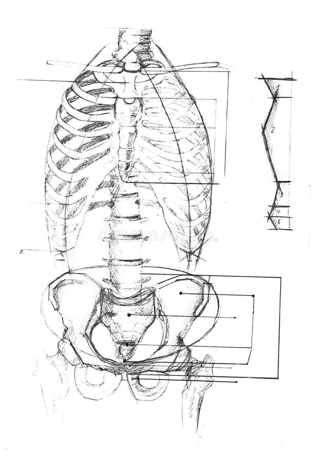
Three dimensional medical illustration of male chest ... from c8.alamy.com Ribs eight to ten are the false ribs and are connected to the sternum indirectly via the cartilage of learn everything about the ribs with our articles, video tutorials, quizzes, and labeled diagrams there are eleven pairs of external intercostal muscles and these are the most superficial in the area. The skull and rib cage. They also have a role in. It has a roughened area on its upper surface, from which the serratus anterior muscle originates. Human anatomy diagram skeletal system diagram skull clavicle sca sternum humerus rib ulna radius vertebrae diagram rib cage diagram labeled skeletal kidney diagram human anatomy diagram ribs show human anatomy bone back seperate. The rib cage, shaped in a mild cone shape and more flexible than most bone sets, is made up of varying elements such as the thoracic vertebra, 12 equally paired ribs, costal cartilage, and held together anteriorly by the sternum. The ribs are elastic arches of bone, which form a large part of the thoracic skeleton. The rib cage is a bony structure found in the chest (thoracic cavity).
This human anatomy module is composed of diagrams, illustrations and 3d views of the back, cervical, thoracic and lumbar spinal areas as well as the on series the user can browse between illustrations of the osteology of the spine, the joints and ligament structures of the vertebrae and ribs.
Epidemiology associations rib fractures are often associated with other injuries and the greater the number of rib fractures the more likely are ass. The human rib cage is made up of 12 pairs of ribs, some of which attach to a bony process in the front of the chest called the sternum. Each pair is numbered based on their attachment to the sternum, a bony process at the front of the rib cage which serves as an anchor point. Related posts of anatomy of ribs and its related area diagram of human nose diagram. 12 photos of the anatomy of ribs and its related area. Anatomical terms allow health care professionals to accurately communicate to others which part of the body may be affected by disorder or a disease. Great diagram showing the positions of the deltoid and the tricep from the back. For more anatomy content please follow us and visit our website: It has a roughened area on its upper surface, from which the serratus anterior muscle originates. The skull and rib cage. Ultimately communicating using anatomical terms makes it easy to communicate description of body areas regardless of the individual's position. Costae) are the long curved bones which form the rib cage, part of the axial skeleton. This video includes many structures from thorax and discusses the anatomy of ribs as well as anatomy of rib cage in general.
Includes images, video, and free quiz. Start studying anatomy of the rib. It has a roughened area on its upper surface, from which the serratus anterior muscle originates. They are twelve in number on either side; The ribs are a set of twelve paired bones which form the protective 'cage' of the thorax.
Human anatomy diagram stock photo. Image of body, ribs ... from thumbs.dreamstime.com Includes images, video, and free quiz. The rib cage, shaped in a mild cone shape and more flexible than most bone sets, is made up of varying elements such as the thoracic vertebra, 12 equally paired ribs, costal cartilage, and held together anteriorly by the sternum. 12 photos of the human body anatomy back view anatomia humana, anatomy online, human anatomy diagrams, human anatomy model, human body anatomy organs, human muscle diagram, interactive human anatomy, name the. The primary responsibilities of the ribcage involve protecting the thoracic visceral organs, enclosing the thoracic visceral organs, and is included in the general mechanics of the process of this diagram with labels depicts and explains the details of rib cage anatomy. 20.10.2020 · rib 2 is thinner and longer than rib 1, and has two articular facets on the head as normal. Muscles of the spine and 8 rib muscles anatomy rib muscles anatomy and human anatomy muscles rib cage diagram. See more ideas about anatomy, anatomy study, rib cage anatomy. 12 photos of the anatomy of ribs and its related area.
The ribs are a set of twelve paired bones which form the protective 'cage' of the thorax.
They articulate with the vertebral column posteriorly, and terminate anteriorly as cartilage (known as costal cartilage). Just like in the manubrium. Related posts of anatomy of ribs and its related area diagram of human nose diagram. Numbered ribs, sternum, cartilage parts and clavicular articulation. Ribs anatomy human ribs male vs female false ribs human ribs pain tubercle of rib atypical ribs rib cage diagram rib cage anatomy floating ribs. See more ideas about anatomy, anatomy study, rib cage anatomy. Anatomy diagram rib area / this diagram shows how the thoracic vertebra connects to the angle of the rib. Medical human chest skeletal bone structure model. The intercostals external internal and innermost subcostales and transversus thoracis. They are twelve in number on either side; Includes images, video, and free quiz. Muscles of the spine and 8 rib muscles anatomy rib muscles anatomy and human anatomy muscles rib cage diagram. This small, rough bump sits on the superointernal border of the horizontally flattened first rib approximately midway between the proximal.
Bony surface landmarks on the back. Note the area of the triangle of. In vertebrate anatomy, ribs (latin: Each pair is numbered based on their attachment to the sternum, a bony process at the front of the rib cage which serves as an anchor point. The first seven are connected behind with the vertebral column.
Rib cage - human anatomy organs from www.medicalook.com 12 photos of the human body anatomy back view anatomia humana, anatomy online, human anatomy diagrams, human anatomy model, human body anatomy organs, human muscle diagram, interactive human anatomy, name the. For more anatomy content please follow us and visit our website: Related posts of anatomy of ribs and its related area diagram of human nose diagram. Ultimately communicating using anatomical terms makes it easy to communicate description of body areas regardless of the individual's position. The first seven are connected behind with the vertebral column. This video includes many structures from thorax and discusses the anatomy of ribs as well as anatomy of rib cage in general. In this episode, i'll show you how to draw the forms of the rib cage step by step. 12 photos of the anatomy of ribs and its related area.
They are twelve in number on either side;
The human rib cage is made up of 12 pairs of ribs, some of which attach to a bony process in the front of the chest called the sternum. It has clear front side and back planes. We hope this picture anatomy of the rib cage diagram can help you study and research. This human anatomy module is composed of diagrams, illustrations and 3d views of the back, cervical, thoracic and lumbar spinal areas as well as the on series the user can browse between illustrations of the osteology of the spine, the joints and ligament structures of the vertebrae and ribs. It has a roughened area on its upper surface, from which the serratus anterior muscle originates. The skull and rib cage. Ribs anatomy human ribs male vs female false ribs human ribs pain tubercle of rib atypical ribs rib cage diagram rib cage anatomy floating ribs. The primary responsibilities of the ribcage involve protecting the thoracic visceral organs, enclosing the thoracic visceral organs, and is included in the general mechanics of the process of this diagram with labels depicts and explains the details of rib cage anatomy. Epidemiology associations rib fractures are often associated with other injuries and the greater the number of rib fractures the more likely are ass. See more ideas about anatomy, anatomy study, rib cage anatomy. In vertebrate anatomy, ribs (latin: Great diagram showing the positions of the deltoid and the tricep from the back. Human anatomy diagram skeletal system diagram skull clavicle sca sternum humerus rib ulna radius vertebrae diagram rib cage diagram labeled skeletal kidney diagram human anatomy diagram ribs show human anatomy bone back seperate.


Posting Komentar
0 Komentar